Persistent Lower Limb Pain Despite Multiple Therapies: A Case Study in Reassessment and Clinical Decision-Making
Not every case follows a straightforward path. This observational case study demonstrates how ongoing reassessment, critical clinical reasoning, and timely referral can be just as important as symptom improvement in managing persistent lower limb pain.
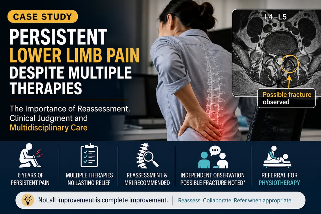
Case Study: Persistent Lower Limb Pain Despite Multiple Therapies — The Importance of Reassessment, Clinical Judgment, and Multidisciplinary Care
Background
Chronic musculoskeletal pain can be challenging to diagnose and manage, particularly when symptoms persist despite prolonged conservative treatment. While many patients improve with non-surgical care, others may experience only partial relief or fail to respond despite multiple interventions.
This case highlights the importance of continuous reassessment, recognising the limitations of a treatment approach, appropriately utilising advanced imaging, and supporting multidisciplinary care when progress is unsatisfactory.
Patient Presentation
A woman in her mid-thirties working in a desk-based occupation presented with a six-year history of persistent lower limb pain that had significantly worsened over the previous one to two months.
The patient reported that symptoms initially began around the anterior aspect of the knee before gradually progressing over time. In an effort to improve, she had pursued multiple conservative strategies including yoga, physiotherapy, supervised gym training, and an extensive course of chiropractic care elsewhere.
Despite these interventions, symptoms persisted and reportedly intensified following periods of increased physical loading during exercise.
At presentation, pain severity was reported as approximately 7–8 out of 10 on most days and was accompanied by bilateral pins and needles sensations involving both lower limbs. The patient otherwise reported good general health with no significant systemic illness.
Previous Management History
Prior to attending our clinic, the patient had undergone numerous conservative interventions without achieving satisfactory long-term improvement.
Despite prolonged treatment and lifestyle modifications, pain continued to interfere with daily activities and quality of life.
This extensive treatment history emphasised the complexity of the presentation and the need for careful reassessment rather than assumptions based solely on previous diagnoses.
Initial Clinical Assessment
Physical examination identified marked muscular hypertonicity and tenderness involving both quadriceps muscles. Even gentle palpation reproduced discomfort, and the patient reported bilateral pins and needles affecting both lower limbs.
Following the initial phase of conservative management, the patient reported noticeable improvement in walking tolerance and complete resolution of the bilateral pins and needles symptoms.
However, despite these encouraging neurological changes, the principal pain complaint remained persistent and continued to significantly affect function.
Repeated examination also identified markedly reduced lumbar segmental mobility that appeared disproportionate to the remainder of the clinical presentation, prompting reconsideration of the working clinical impression.
Given the prolonged history, persistent pain, and atypical response pattern, further investigation was recommended.
Advanced Imaging and Clinical Observation
Magnetic resonance imaging (MRI) and orthopaedic assessment were subsequently obtained.
According to the patient, the formal imaging review and specialist consultation did not identify a definitive structural explanation for the symptoms.
As part of routine clinical assessment, the treating clinician independently reviewed the MRI images. During this review, an imaging feature involving the posterior elements at the L4–L5 level raised consideration of a possible structural abnormality, including the possibility of a fracture.
However, this observation was not confirmed by the available radiology report or orthopaedic assessment. Consequently, no definitive diagnosis was established, and no management decisions were based solely on this independent interpretation.
Instead, the imaging review reinforced the importance of maintaining diagnostic caution and integrating imaging findings with the patient's history, examination findings, and specialist input.
Management and Clinical Decision-Making
Conservative care continued using symptom-guided manual therapy, supportive modalities, movement advice, and rehabilitation principles.
Although neurological-type symptoms improved and mobility became more comfortable, the patient's primary pain remained severe and functionally limiting.
Repeated reassessment suggested that further continuation of the existing management approach alone was unlikely to achieve satisfactory progress.
In the interest of patient-centred care, a multidisciplinary pathway was recommended. The patient was advised to continue rehabilitation through physiotherapy, and detailed clinical notes outlining examination findings and observations were provided to support continuity of care.
This recommendation reflected the clinical judgment that referral and collaborative management were more appropriate than simply extending conservative treatment without meaningful overall improvement.
Clinical Reflection
One of the most important responsibilities in conservative musculoskeletal practice is recognising when treatment goals are not being met.
While symptom improvement is encouraging, partial improvement should not be mistaken for complete resolution, particularly when significant pain and disability remain.
This case demonstrates the value of reassessing assumptions, recommending further investigation when appropriate, and being prepared to modify or discontinue a treatment pathway when progress is insufficient.
Equally important, independent observations made during image review should be interpreted cautiously and should not replace formal radiological interpretation or specialist evaluation when confirmation is lacking.
Key Clinical Lessons
Chronic pain presentations often require repeated reassessment and flexible clinical reasoning.
Improvement in one symptom domain does not necessarily indicate resolution of the underlying condition.
Imaging findings should always be interpreted within the context of formal radiology reporting, specialist opinion, and overall clinical assessment.
Unconfirmed imaging observations should remain clinical considerations rather than definitive diagnoses.
Appropriate referral and multidisciplinary collaboration are important aspects of responsible patient care.
Recognising when continued treatment is unlikely to provide meaningful additional benefit is an essential component of ethical clinical practice.
Important Disclaimer
This article presents a single anonymised observational clinical case for educational purposes.
Clinical details have been modified where appropriate to protect patient privacy.
The MRI observation described represents an independent clinical interpretation that was not confirmed by the available radiology report or orthopaedic assessment. It should therefore not be interpreted as a confirmed fracture or established structural diagnosis.
This report does not establish causation or treatment effectiveness and should not be interpreted as medical advice. Individual presentations and outcomes vary, and persistent or worsening symptoms should be evaluated by an appropriately qualified healthcare professional.